The PennHIP method is a novel way to assess, measure and interpret hip joint laxity. It consists of three separate radiographs: the distraction view, the compression view and the hip extended view.
It is the amount of displacement of the femoral head from the acetabulum during distraction radiography that has been termed passive hip laxity and that has been shown to be directly related to the probability that a hip will develop degenerative joint disease characteristic of hip dysplasia. The distraction view and compression view, are used to obtain accurate and precise measurements of joint laxity and congruity. The hip-extended view is used to obtain supplementary information regarding the existence of degenerative joint disease (DJD) of the hip joint. (The hip-extended view is the conventional radiographic view used to evaluate the integrity of the canine hip joint.)
The PennHIP technique is more accurate than the current standard and it has been shown to be a better predictor for the onset of DJD. The radiographs below are of the same dog, yet the hip joint laxties in each view look very different. Notice that the hips in the distraction view appear to be much looser than they do in the hip-extended view.
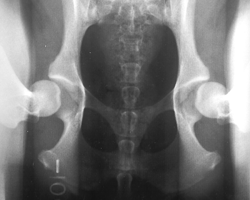
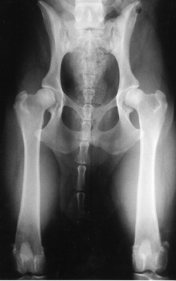
The obvious contrast in joint laxity between the distraction and hip-extended views demonstrates the fundamental difference between the two radiographs. The looser the joint on the distraction view, the greater is the chance that the hip will develop DJD. The hip-extended view tends to mask true hip joint laxity because the joint capsule is wound up into a tightened orientation when the hips are extended. This explains why measurable joint laxity on the distraction view is always greater than the measurable laxity from the hip-extended view. In fact, distraction laxity is up to 11 times greater depending on the breed of dog under study.
The compression view is used to determine the “goodness of fit” of the femoral heads into the acetabula. In a hip with DJD, the remodeling that occurs in the acetabulum and/or the femoral head, will often result in an ill-fitting “ball” and “socket” (see Primer of Canine Hip dysplasia for an example of DJD).To summarize, the PennHIP method:
– Obtains DJD readings from the standard hip-extended view
– Obtains hip joint congruity readings from the compression view
– Obtains quantitative measurements of hip joint laxity from the distraction view
Why do the hips appear looser on the distraction view?
For a brief review of the anatomy of the canine hip joint, see the Primer on Canine hip dylsplasia article.
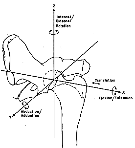
A ball and socket joint has the ability to rotate about the three orthogonal (XYZ) axes. With respect to the hip, the femur can rotate back and forth (flexion/extension), medial and lateral (adduction/abduction), and can rotate “toe in”/”toe out” (internal/external rotation). The diagram at the right illustrates these points. Palpation of the hip also demonstrates that the femoral head can be translated (displaced) laterally. That is, it moves out or away from the acetabular cup. This lateral displacement, although an important property of the hip joint, had not been adequately studied prior to 1983. The amount of the displacement is simply the laxity of the joint.The maximum amount of displacement of the femoral head in the neutral position is not limited by the length of the round ligament as once thought. Instead, it is dependent on the relative volume of the synovial fluid in the joint, acting in combination with the joint capsule.
Biomechanical testing in the laboratory showed that femoral head displacement is greatest when the femur is in a position approximating a neutral or “standing” position, also called the stance-phase of weight bearing. The legs are placed in this position for the distraction view.In the familiar hip-extended view, the femur is pulled into extension (see the diagram at left). The fibrous elements of the joint capsule get “wound up” so to speak, so that the resulting tension serves to drive the femoral head into the acetabulum (socket). This explains why the measurable joint laxity on the distraction view is always greater than the measurable laxity from the hip-extended view.It follows that a loose-hipped dog, such as the one at the beginning of this page, could falsely be considered to have “tight” or “normal” hips according to the hip-extended radiograph. Such a dog would likely be approved for breeding and therefore could pass this loose-hipped trait onto its offspring.
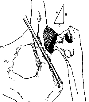
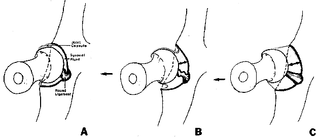
In the schematic below, figure A depicts a normal joint in the neutral position. The joint capsule and the round ligament are lax (relaxed, but not stretched) when there is no distractive force being applied. When a force is applied (figure B) the joint capsule invaginates reflecting a pressure differential (vacuum effect) across the joint capsule. The relative degree of displacement is dependent upon the amount of synovial fluid present. More synovial fluid volume means greater hip laxity. Notice that the round ligament remains lax in this position. In a dog with excessive synovial fluid volume (figure C) the capsule does not invaginate and the magnitude of femoral head displacement is limited by the the joint capsule, the surrounding musculature and the round ligament. The distraction radiographic procedure shows the relative displacement of the femoral head from the acetabulum when a distractive force is applied, however the synovial fluid and joint capsule are transparent to x-rays and therefore are not visible on the radiograph. It is the amount of displacement of the femoral head from the acetabulum during distraction radiography that has been termed “passive hip laxity” and that has been shown to be directly related to the probability that a hip will develop degenerative joint disease characteristic of hip dysplasia.
Comments are closed.